Dow v. Miller: Medical Legal Visualization
Client
Dr. Leila Lax, Biomedical Communications, University of TorontoTeam members
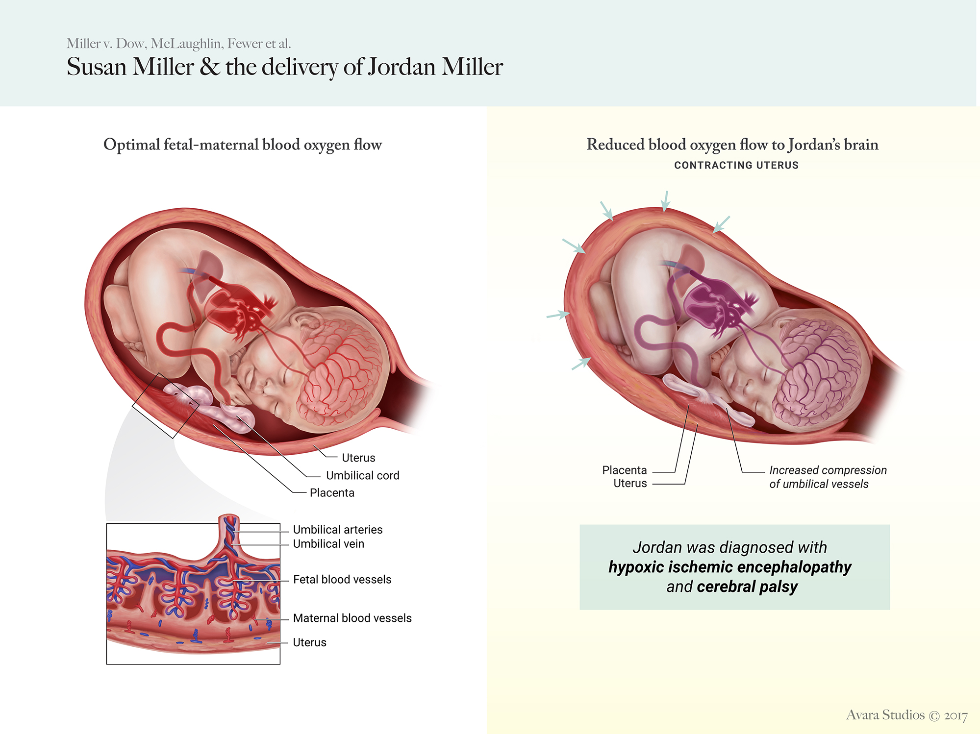
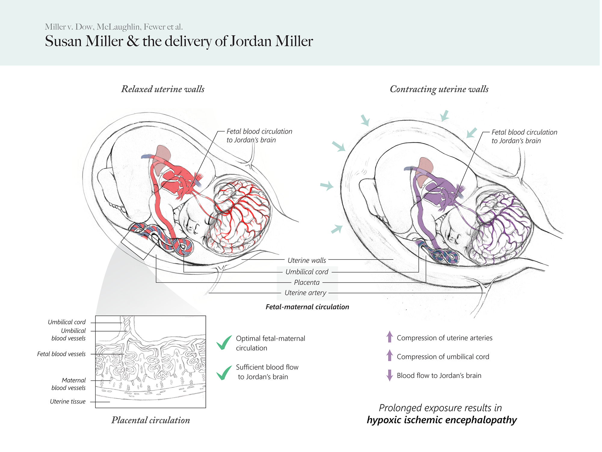
Sarah CrawleyThe two medical legal visualization panels are intended for plaintiff use as demonstrative evidence in the Canadian courtroom. The plaintiffs (Susan Miller et. al) argued that considering Susan Miller's situation in her labour and delivery of her son, Jordan Miller, it was crucial to include fetal electrode monitoring. Therefore, Dr. Dow and his team's failure to include internal monitoring lead to their inability to detect fetal distress thus allowing Jordan to suffer from hypoxic ischemia and ultimately, cerebral palsy.
Audience
Canadian judge and juryMedia
Adobe Photoshop, Adobe IllustratorDate completed
December 2017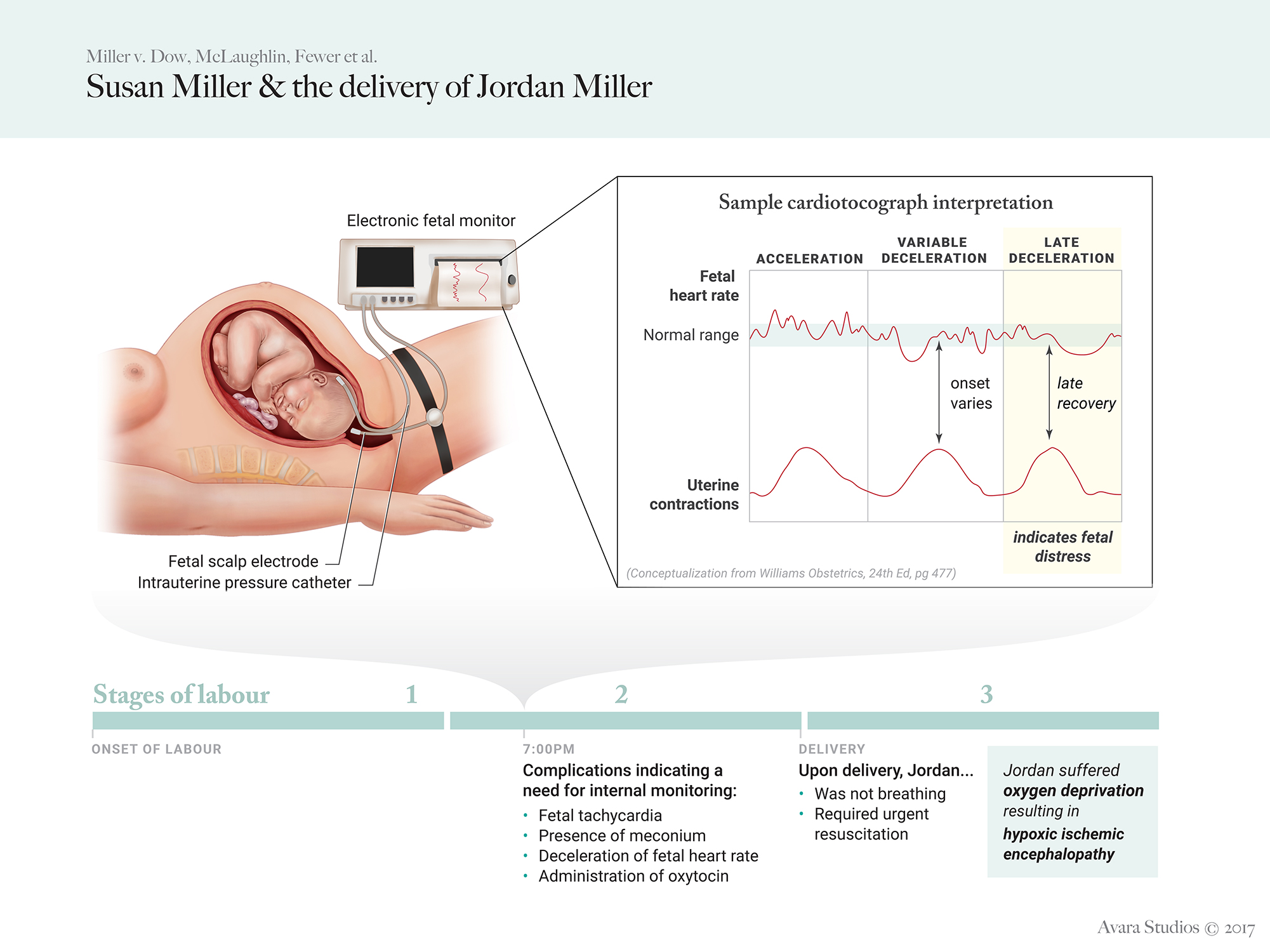
Process
Research
We were given a file describing the Dow v. Miller case. Due to lack of monitoring, there were no readings, graphs or any quantitative data to be working from, therefore, we based all of our visualizations on expert witness testimonies from the case file and our own research.
Our biggest challenge was trying to visualize the science, despite the huge gaps of information in the case, while also being mindful not to imply more than what was presented in the case and bias the narrative.
Co-visualization
Sarah and I worked closely together to come up with the initial conceptualization. In order to maintain visual consistency, we developed a style sheet and a fair render plan that would allow for us to work off of each other's illustrations. We were constantly brainstorming together, providing mutual feedback and making detailed edits to maintain an accurate visualization and cohesive visual style.


Works consulted
Cunningham, F. G., Leveno, J. K., Bloom L. S., Spong, Y. C., Dashe, S. J, Hoffman, L. B., Casey, M. B. and Sheffield, S. J (Ed.). (2014). Williams Obstetrics: 24th Edition. New York : McGraw-Hill Education/Medical.
Edmonds, D. K. (2007). Dewhurst's textbook of obstetrics & gynaecology: 7th Edition. Malden, Massachusetts: Wiley-Blackwell Publishers.
Hutter, D., Kingdom, J. and Jaeggi, E. Causes and Mechanisms of Intrauterine Hypoxia and Its Impact on the Fetal Cardiovascular System: A Review. International Journal of Pediatrics: 2010. DOI:10.1155/2010/401323.
RSD-K3 10.4” fetal monitor. Shenzhen Setolink Electronics Co., Ltd. [Product image]. Available at: https://www.alibaba.com/product-detail/HIGH-QUALITY-AND-LOW-PRICE-10_60289081842.html.
Uflacker, Renan. (2007). Atlas of vascular anatomy: an angiographic approach: 2nd Edition. Philadelphia : Lippincott Williams & Wilkins.
Zia, Shumaila. (2013). Placental location and pregnancy outcome. Journal Of The Turkish-German Gynecological Association, 14: 190-3.
Nucleus Medical Media. Normal Uterine Contractions During Delivery with Fetal Monitoring Strip. Available at: http://www.nucleuscatalog.com/normal-uterine-contractions-during-delivery-with-fetal-monitoring-strip/view-item?ItemID=36700.
Nucleus Medical Media. Placental Abruption with Fetal Hypoxia. Available at: http://www.nucleuscatalog.com/placental-abruption-with-fetal-hypoxia/view-item?ItemID=12208
Nucleus Medical Media. Hypovolemic and Hypoxic Ischemic Encephalopathy (Brain Damage). Available at: http://www.nucleuscatalog.com/hypovolemic-and-hypoxic-ischemic-encephalopathy-brain-damage/view-item?ItemID=3499.
Femicare: Center of prenatal ultrasonographic diagnostics. (2012). 3D ultrasound - third trimester of pregnancy (photoseries). Available at: http://www.femicare.org/en/3d-4d-ultrasound.